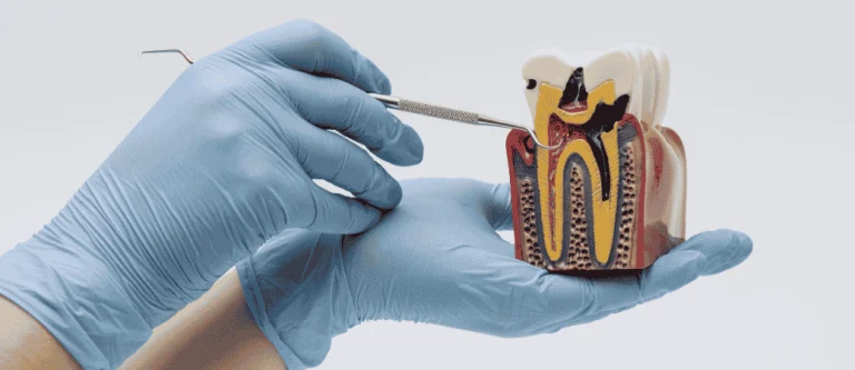
A tooth can look healthy on the outside while inflammation or infection develops around its roots.
AI root canal infection detection is an emerging approach that uses artificial intelligence to help dentists analyze dental images and identify areas that may need closer examination.
These tools can highlight suspicious patterns, but they cannot confirm an infection without a clinical evaluation.
These tools analyze dental radiographs for patterns associated with changes around the root tip. Some changes may appear without noticeable symptoms, although AI cannot reliably identify every early infection.
This doesn’t mean a computer is diagnosing your tooth. AI software flags areas worth a second look; your dentist still confirms what’s actually happening and decides what to do next.
Hidden root infections matter because they can sit quietly for months. By the time a tooth throbs or swells, the infection may have already caused measurable bone loss around the root. Catching subtle clues earlier gives you more treatment options and may mean a simpler visit.
Key Takeaways
- AI tools can flag faint bone changes near a tooth root that may be easy to miss on a routine X-ray.
- Periapical X-rays and CBCT scans serve different purposes, and AI can analyze both, though CBCT adds detail that isn’t always necessary.
- An AI flag is a starting point for your dentist’s exam, not a finished diagnosis or a treatment decision on its own.
How Can AI Spot a Root Canal Infection That Is Easy to Miss?
AI software trained on dental X-rays can pick out small shadows and bone changes near a root tip that signal early apical periodontitis, a condition where the tissue around the root becomes inflamed or infected.
These programs compare patterns across thousands of images, which helps them notice subtle density changes a tired eye might skip over during a busy day of chart reviews.
This matters because early periapical lesions don’t always come with pain. A tooth can harbor a slow, quiet infection for a long stretch before any symptom shows up, which is part of why endodontic disease diagnosis has always depended heavily on image quality and a sharp eye.
Research reviewing AI tools in endodontics has pointed to improved diagnostic accuracy compared with unaided visual reading in some study settings.
What Subtle Changes Appear Around a Tooth’s Root?
A root with a brewing infection often shows a faint widening of the space between the tooth and surrounding bone, or a small dark patch near the root tip.
These changes can look like background noise on a standard radiograph, especially if the lesion is still small.
Some AI models are trained to identify radiographic patterns associated with periapical lesions. Their ability to detect subtle changes depends on image quality, the training data, and the specific software used.
Can AI Find a Lesion Before Symptoms Become Severe?
AI tools may flag early-stage changes around a root before a tooth becomes painful or sensitive to temperature. This gives your dentist a chance to monitor or treat a problem while it’s still small and simpler to manage.
A flagged image still needs clinical judgment. Your dentist will look at your symptoms, test the tooth, and decide whether more follow-up is needed before suggesting root canal treatment or another next step.
Which Dental Images Can AI Analyze?
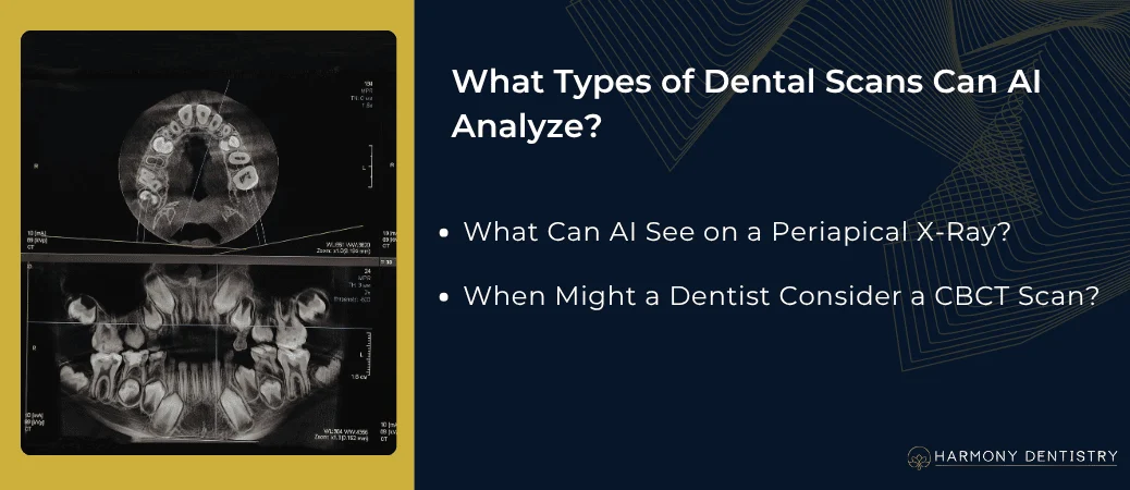
AI software can review the same images your dentist already takes, including periapical radiographs, bitewings, panoramic films, and cone-beam computed tomography scans.
Each image type gives a different level of detail, and AI adapts its analysis to what that specific format can show.
Routine dental imaging remains the first stop for most checkups, since it’s fast and uses a low dose of radiation. CBCT adds a three-dimensional view when a tooth’s anatomy or a suspected infection needs a closer look.
Learn more about the dental technology used at Harmony Dentistry to support patient evaluations and treatment planning.
What Can AI See on a Periapical X-Ray?
A periapical X-ray gives AI software a flat, two-dimensional view of the tooth root and surrounding bone.
AI can scan this image for early signs of bone change, root fracture patterns, and shifts in the shape of the space around the root tip.
This type of film works well for routine screening and catches many problems early. Its flat view can sometimes hide a lesion that sits directly behind or in front of a root, which is one reason dentists sometimes need more detail.
When Might a Dentist Consider a CBCT Scan?
A dentist may suggest a CBCT scan when a periapical X-ray doesn’t match the symptoms you’re describing, or when a tooth’s root anatomy looks unusually complex.
CBCT builds a 3D image, letting AI and your dentist view a root from every angle instead of just one flat plane.
This added detail can reveal small lesions hidden behind the root or tucked between roots that a flat film might miss entirely.
Research on AI-assisted CBCT analysis has pointed to improved detection of periapical lesions and complex root canal shapes, as shown in a study on AI-driven diagnostic accuracy with CBCT data.
A CBCT scan isn’t automatic for every visit; it carries a higher radiation dose than a standard film, so dentists reserve it for cases where the extra detail is likely to change the plan.
What Else Can AI Reveal About a Difficult Tooth?
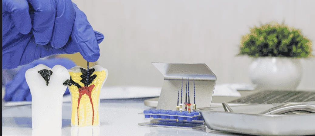
AI can help map out root canal morphology, including canals that are easy to overlook during treatment planning.
This matters most in teeth known for complicated internal shapes, where missing a canal can leave infection behind even after a root canal is finished.
Maxillary molars are a common example. These back upper teeth often carry an extra canal called the mb2 canal, which can be narrow, curved, or positioned in a spot that’s hard to spot on a flat image.
Can AI Help Identify a Missed Canal?
AI software trained on CBCT and radiograph data can highlight likely canal locations, including extra canals that standard imaging sometimes hides. This can give your dentist a clearer map before or during treatment, lowering the chance a canal gets missed.
A missed canal can let infection linger even after a root canal seems complete. Catching dental anomalies like an extra canal early can mean one successful visit instead of a need for retreatment later.
Can It Distinguish Root Changes from Nearby Bone Loss?
AI tools are trained to separate changes tied to the root tip from bone loss caused by periodontal disease, which affects the gums and supporting bone along the side of a tooth.
These two problems can look similar on an X-ray but need very different treatment.
Telling them apart matters because periodontal bone loss calls for gum-focused care, while a root tip lesion often points toward root canal treatment.
A dentist uses the shape, location, and pattern of bone loss to tell the two apart, with AI serving as an extra set of eyes rather than a final word.
How Reliable Are AI Findings?
AI accuracy depends heavily on how a given model was built and trained, which means published numbers reflect specific study conditions rather than a guarantee for every dental office.
Several types of models power today’s dental AI tools, including convolutional neural networks, resnet, efficientnet, u-net, and newer vision transformer designs.
These names describe different ways a program learns to recognize patterns in an image. Each has strengths; some are better at drawing an outline around a lesion, while others are tuned for classifying an image as showing disease or not.
How Do Image-Analysis Models Learn to Recognize Problems?
Machine learning and deep learning models learn by studying thousands of labeled X-rays where a dentist has already marked where disease appears.
Over time, the model adjusts internal settings until it can spot similar patterns on new, unlabeled images.
Many dental AI tools use transfer learning, which means they start from a model already trained on a large general image set, then get fine-tuned on dental radiographs.
Some systems include a tool called grad-cam, which creates a heat map showing which part of the image led the model to its conclusion, giving dentists a way to check the AI’s reasoning rather than trusting it blindly.
Why Can Results Vary Between Studies and Dental Offices?
Accuracy numbers from one study don’t always carry over to a different office or patient group.
Differences in X-ray equipment, image quality, patient population, and even how a lesion was defined in the training data can all shift results.
What Happens After AI Flags a Possible Infection?
An AI alert is not a confirmed diagnosis. At Harmony Dentistry, a dentist evaluates symptoms, dental history, clinical findings, and appropriate imaging before recommending treatment.
AI-assisted image analysis, where available, may provide additional information, but clinical judgment remains essential.
This step matters because an X-ray shadow can come from several sources, including pulpitis (inflammation inside the tooth’s pulp) or other endodontic diseases that don’t always need the same treatment.
A flagged image tells your dentist where to look closer, not what to do next.
When a tooth cannot be predictably restored, tooth extraction may be considered after a clinical evaluation.
How Does a Dentist Confirm What the Image Suggests?
A dentist confirms a suspected infection through a combination of tests, including checking how the tooth responds to cold or heat, tapping on it to check for tenderness, and reviewing your reported symptoms.
These clinical findings get compared against what the image shows before any treatment decision gets made.
This step can’t be skipped. An image alone can’t tell your dentist whether a tooth’s nerve is still alive, which is a key piece of information when deciding between watching a tooth, treating it, or referring you for a specialist opinion.
Does an AI Alert Always Mean Root Canal Treatment?
An AI alert does not always mean you need root canal treatment. Some flagged areas turn out to be early, reversible inflammation that can settle with simpler care, while others may need monitoring with a follow-up X-ray before any procedure gets scheduled.
Your dentist weighs the image alongside your symptoms and exam findings before recommending root canal treatment or another path.
If you’re unsure what a recommendation means for your specific tooth, the patient resources section covers what to expect at different stages of care.
Earlier Clues Support Better-Informed Care
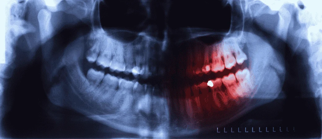
AI’s growing role in reading dental images gives dentists an extra layer of support when symptoms and X-rays don’t quite match up.
It can flag faint bone changes, highlight a possible missed canal, and point toward complex root anatomy that calls for a closer look.
None of this replaces a dentist’s exam, your reported symptoms, or the judgment built from years of clinical training.
AI works best as a second set of eyes, helping surface the images worth a second look so nothing slips through during a routine visit.
If you are wondering how regular dental visits can help identify problems before they become more serious, read about the importance of biannual dental visits and how routine checkups can catch dental issues early.
Frequently Asked Questions
Can AI detect a root canal infection before it hurts?
AI software can flag early bone changes around a root tip before a tooth becomes painful, since infection often shows on an image before symptoms appear.
A dentist still needs to confirm the finding with clinical tests, since an early flag doesn’t always mean treatment is needed right away.
How accurate is AI at finding infections on dental X-rays?
Accuracy varies by study, by the type of AI model used, and by the quality of the images it was trained on.
Published figures describe performance in specific research settings and don’t guarantee the same results in every dental office.
Do I need a CBCT scan to find a hidden root canal infection?
Not always. A standard periapical X-ray catches many infections just fine, and a CBCT scan gets reserved for cases where symptoms and the regular X-ray don’t match up, or when root anatomy looks unusually complex.
Can AI tell whether I need a root canal?
AI can highlight a suspicious area on an image, but it can’t decide on its own whether you need a root canal. That decision depends on your symptoms, a clinical exam, and your dentist’s judgment, with the AI flag serving as one more piece of information.
Will AI replace dentists or endodontists?
AI is a support tool, not a replacement for a trained dentist or endodontist. It helps highlight patterns on images, while diagnosis and treatment planning still depend on clinical exams, patient history, and hands-on professional judgment.